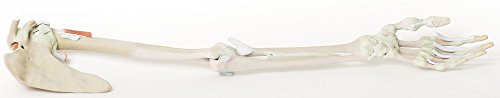
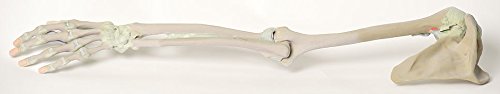
At the forefront of medicine and technology, we are proud to offer these incredible, uncompromised replicas of human anatomy. Using the latest 3D printing technology and materials available, this model is an exact replica of a human cadaver, brought to "life" by extensive medical scanning and manufacturing technologies. Over are the days of using ethically questionable cadavers, the mess of hazardous preservation chemicals, and the inaccuracies of plastinated models that often over-enhance anatomy for display, not realism. See the future, and the beauty, of real human anatomy with these incredible anatomical replicas! This 3D printed specimen presents the entire upper limb skeleton and ligaments from the pectoral girdle to the hand. In the pectoral girdle, the ligaments spanning the clavicle and scapula (acromioclavicular, coracoclavicular, coracoacromial) as well as the superior transverse scapular ligament spanning the suprascapular notch, are visible. A small portion of the supraspinatus muscle belly and tendon are preserved to demonstrate the passage of the muscle deep to the coracoacromial ligament, which is a very clinically relevant area of anatomy. The tendon of the subscapularis muscle has been reflected slightly to expose the anterior aspect of the glenohumeral joint capsule, and the tendon of the long head of triceps brachii, teres major, and latissimus dorsi are preserved surrounding the capsule and proximal humerus. The tendon of the long head of biceps brachii is visible within the intertubercular groove, and exposed within the superior glenohumeral joint capsule as it approaches the supraglenoid tubercle. The capsule of the elbow joint has been dissected to expose the articular surfaces of the distal humerus, proximal radius and proximal ulna. Both the ulnar and radial collateral ligaments are preserved, as is the annular ligament of the radius. Just distal to the joint capsule, the tendinous insertion of the biceps brachii is preserved as it inse
Trustpilot
2 weeks ago
4 days ago
