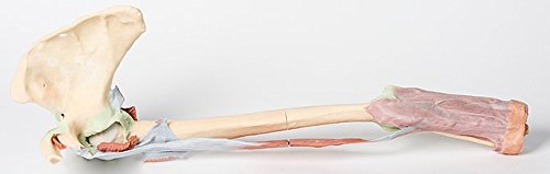
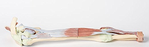
At the forefront of medicine and technology, we are proud to offer these incredible, uncompromised replicas of human anatomy. Using the latest 3D printing technology and materials available, this model is an exact replica of a human cadaver, brought to "life" by extensive medical scanning and manufacturing technologies. Over are the days of using ethically questionable cadavers, the mess of hazardous preservation chemicals, and the inaccuracies of plastinated models that often over-enhance anatomy for display, not realism. See the future, and the beauty, of real human anatomy with these incredible anatomical replicas! This 3D print shows the origin and insertion of biceps (most other arm and shoulder muscle bellies have been removed). The long head of biceps arises from the supraglenoid tubercle (hidden from view) and travels inferiorly in the bicipital groove, whereas the short head of biceps arises from the coracoid process. The bifid insertion of the muscle as the bicipital aponeurosis and the rounded tendon which can be seen winding around the radius to insert into the radial tuberosity are clearly discernable. At the shoulder region the dissected attachments of some muscles (subclavius, subscapularis, pectoralis major, teres minor, infraspinatus, long head of triceps) and the tendinous insertion of latissimus dorsi can be identified close to the 'floor' of the medial lip of the bicipital groove. The tendon of teres major lies on the medial lip of the groove and the pectoralis major tendon inserts into the lateral lip of the groove. The tendon of pectoralis minor arises from the coracoid process medial to the origin of the short head of biceps. Ligaments of the shoulder region such as the coracoclavicular, coracoacromial, coracohumeral are visible, as is the glenohumeral and acromioclavicular joint capsules. The supraspinatus muscle is the only rotator cuff muscle that has been completely preserved. The suprascapular ligament which bridges across the supras
Trustpilot
1 month ago
1 day ago
