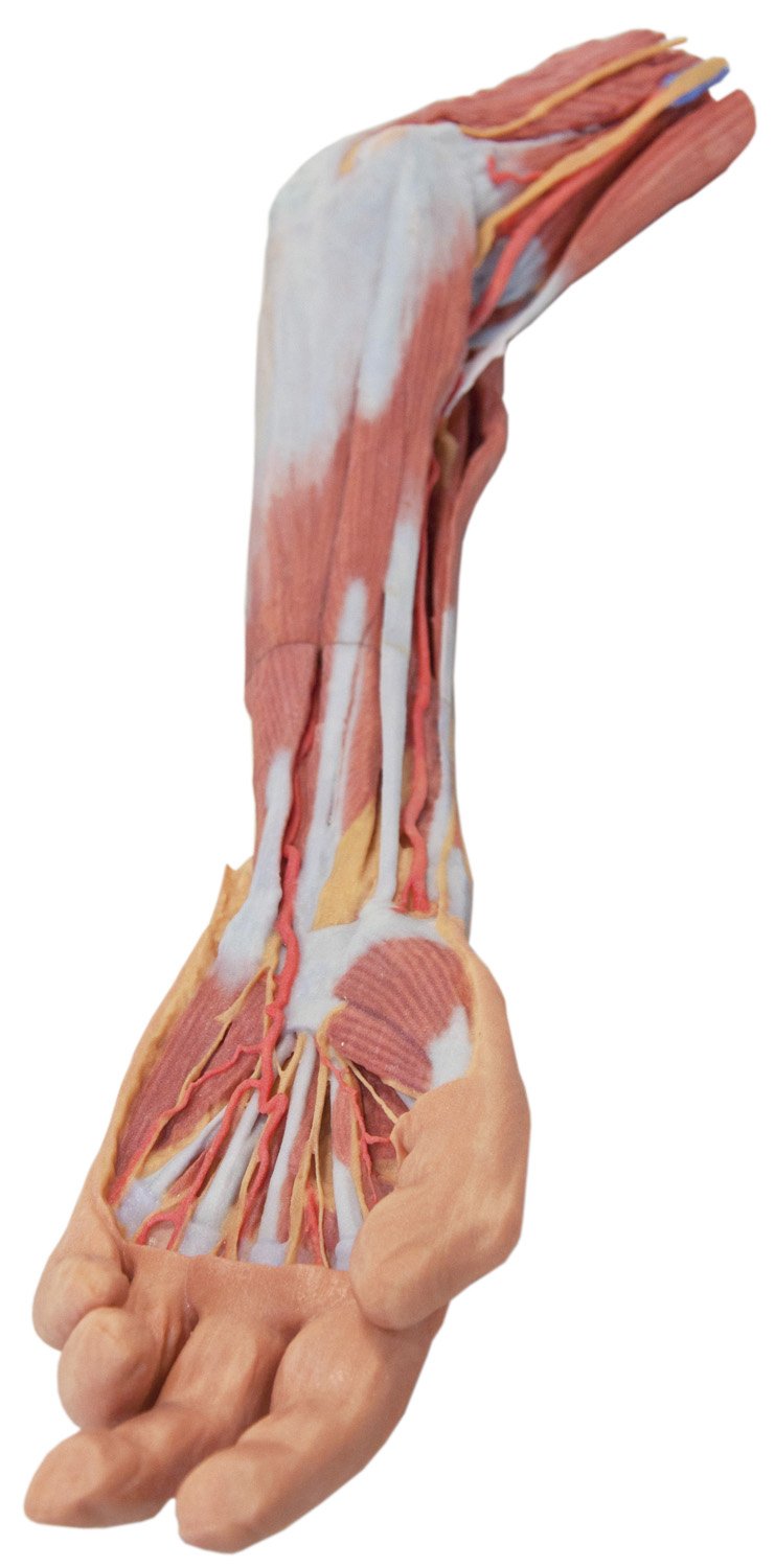


At the forefront of medicine and technology, we are proud to offer these incredible, uncompromised replicas of human anatomy. Using the latest 3D printing technology and materials available, this model is an exact replica of a human cadaver, brought to "life" by extensive medical scanning and manufacturing technologies. Over are the days of using ethically questionable cadavers, the mess of hazardous preservation chemicals, and the inaccuracies of plastinated models that often over-enhance anatomy for display, not realism. See the future, and the beauty, of real human anatomy with these incredible anatomical replicas! This upper limb specimen displays the vascular, nervous and muscular anatomy of a left distal arm, forearm and hand. In the distal arm and elbow/cubital fossa region we can see the arrangement of the biceps tendon, brachial artery and median nerve from lateral to medial. The bicipital aponeurosis has been divided to reveal the structures deep to it. The ulnar nerve can be seen passing behind the medial epicondyle with an ulnar collateral artery close by. The superficial branch of the radial nerve can just be seen in the space between brachioradialis and brachialis muscles (as the belly of the latter muscle has been displaced slightly laterally). In the forearm, the superficial flexor muscles arising from the common flexor origin can be clearly seen (from lateral to medial pronator teres, flexor carpi radialis (FCR), flexor digitorum superficialis (FDS) and flexor carpi ulnaris (FCU)). There is not a palmaris longus muscle in this cadaver. The radial artery and superficial branch of the radial nerve (emerging half way down the forearm from behind the brachioradialis muscle and tendon) are clearly identifiable. The ulnar artery can be seen in the distal forearm emerging from beneath FCU muscle. On the posterior aspect of the forearm the extensor muscles arising from the common extensor origin are clearly identifiable. These include (from medial to
Trustpilot
1 day ago
2 weeks ago
