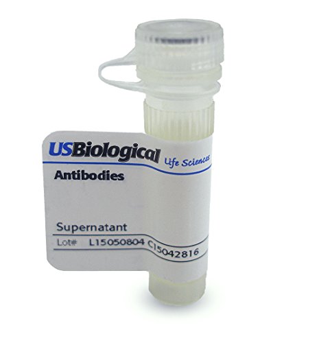

Recognizes a phosphor-protein of 45kDa, identified as MyoD1. The epitope of this MAb maps between amino acid 180-189 in the C-terminal of mouse MyoD1 protein. It does not cross react with myogenin, Myf5, or Myf6. Antibody to MyoD1 labels the nuclei of myoblasts in developing muscle tissues. MyoD1 is not detected in normal adult tissue, but is highly expressed in the tumor cell nuclei of rhabdomyosarcomas. Occasionally nuclear expression of MyoD1 is seen in ectomesenchymoma and a subset of Wilm’s tumors. Weak cytoplasmic staining is observed in several non-muscle tissues, including glandular epithelium and also in rhabdomyosarcomas, neuroblastomas, Ewing’s sarcomas and alveolar soft part sarcomas. Applications:  Suitable for use in Immunofluorescence, Flow Cytometry, Western Blot, Immunoprecipitation, Immunohistochemistry. Other applications not tested. Recommended Dilution: Flow Cytometry: 0.5-1ug/million cells  Immunofluorescence: 0.5-1ug/ml  Western Blot: 0.25-0.5ug/ml  Immunoprecipitation: 0.5-1ug/500ug protein lysate Immunohistochemistry: Frozen & Formalin-fixed: 0.5-1ug/ml for 30 minutes at RT (Staining of formalin-fixed tissues is enhanced by boiling tissue sections in 1mM EDTA Buffer, pH 7.5-8.5, for 10-20 min followed by cooling at RT for 20 minutes Optimal dilutions to be determined by the researcher. Positive Control: Rhabdomyosarcoma Storage and Stability: May be stored at 4°C before opening. DO NOT FREEZE! Stable at 4°C as an undiluted liquid. Dilute only prior to immediate use. Stable for 12 months after receipt. For maximum recovery of product, centrifuge the original vial after thawing and prior to removing the cap. Further dilutions can be made in assay buffer. Freezing alkaline phosphatase conjugates will result in a substantial loss of activity. Note: Applications are based on unconjugated antibody.
Trustpilot
1 month ago
4 days ago
2 months ago
1 month ago